Embark on a comprehensive exploration of concept map muscle structural organization, a meticulous framework that unravels the intricate levels of muscle architecture. From the fundamental building blocks of myofibrils to the intricate organization of muscle fibers and tissues, this concept map provides a comprehensive guide to the structural underpinnings of muscle function.
Delving into the hierarchical levels of muscle organization, we uncover the intricate interplay between myofilaments, sarcomeres, and the sarcoplasmic reticulum, revealing the molecular mechanisms that govern muscle contraction. Moreover, we examine the diverse types of muscle fibers and their specialized functional properties, providing a deeper understanding of muscle diversity and adaptability.
Introduction: Concept Map Muscle Structural Organization

Muscle structural organization refers to the hierarchical arrangement of muscle tissue from the smallest unit to the whole muscle. Understanding this organization is crucial for comprehending muscle function and movement.
The hierarchical levels of muscle organization are:
- Myofibril
- Muscle fiber
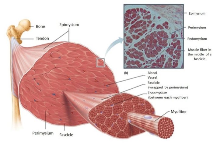
- Muscle fascicle
- Muscle bundle
- Whole muscle
Myofibril Organization
Myofibrils are cylindrical structures within muscle fibers that contain the contractile machinery. They are composed of repeating units called sarcomeres, which are the fundamental units of muscle contraction.
Each sarcomere is made up of thick filaments (myosin) and thin filaments (actin), arranged in a regular pattern. The sliding of these filaments past each other during muscle contraction generates force.
The sarcoplasmic reticulum (SR) and T-tubules play vital roles in muscle contraction. The SR stores calcium ions, which are released into the sarcoplasm to initiate contraction. T-tubules are invaginations of the sarcolemma that allow the rapid spread of action potentials deep into the muscle fiber, triggering the release of calcium ions from the SR.
Muscle Fiber Structure
Muscle fibers are the basic functional units of muscle tissue. They are long, cylindrical cells surrounded by a sarcolemma, the cell membrane of a muscle fiber.
The sarcoplasm is the cytoplasm of a muscle fiber and contains myofilaments (actin and myosin), as well as other cellular components. Myofilaments are arranged in a repeating pattern called myofibrils.
There are different types of muscle fibers, each with distinct functional properties:
- Type I (slow-twitch) fibers: Slow to contract and fatigue, suitable for endurance activities.
- Type IIa (fast-twitch, oxidative-glycolytic) fibers: Intermediate contraction speed and fatigue resistance, used in both endurance and power activities.
- Type IIx (fast-twitch, glycolytic) fibers: Fast to contract but fatigue quickly, specialized for power activities.
Muscle Tissue Organization
Muscle fibers are organized into fascicles, which are bundles of fibers surrounded by a layer of connective tissue called the perimysium.
Fascicles are further organized into muscle bundles, which are larger bundles surrounded by the epimysium, a thicker layer of connective tissue. The epimysium connects the muscle to surrounding structures.
There are three main types of muscle tissue:
- Skeletal muscle: Attached to bones and responsible for voluntary movement.
- Smooth muscle: Found in the walls of internal organs and responsible for involuntary functions such as digestion and blood vessel constriction.
- Cardiac muscle: Found only in the heart and responsible for pumping blood.
Neuromuscular Junction, Concept map muscle structural organization
The neuromuscular junction is the site where a motor neuron communicates with a muscle fiber. It consists of the motor neuron terminal, the synaptic cleft, and acetylcholine receptors on the muscle fiber membrane.
When an action potential reaches the motor neuron terminal, it triggers the release of acetylcholine into the synaptic cleft. Acetylcholine binds to receptors on the muscle fiber membrane, causing the opening of ion channels and the generation of an action potential in the muscle fiber, initiating muscle contraction.
Muscle Contraction
Muscle contraction is the process by which muscles generate force and movement. It involves the sliding of thick and thin filaments past each other within the sarcomere.
The sliding filament theory explains that during contraction, myosin heads bind to actin and undergo a conformational change, pulling the thin filaments towards the center of the sarcomere. This movement shortens the sarcomere and generates force.
Calcium ions play a crucial role in muscle contraction. They bind to troponin on the thin filaments, causing a conformational change that exposes the myosin-binding sites on actin.
Muscle contraction requires energy, which is provided by the breakdown of ATP. The regulation of muscle contraction involves a complex interplay of calcium ions, ATP, and regulatory proteins.
Answers to Common Questions
What is the significance of the sarcoplasmic reticulum in muscle contraction?
The sarcoplasmic reticulum plays a crucial role in muscle contraction by storing and releasing calcium ions. Calcium ions trigger the sliding of myofilaments, initiating the contraction process.
How do different types of muscle fibers contribute to muscle function?
Different types of muscle fibers, such as slow-twitch and fast-twitch fibers, exhibit distinct functional properties. Slow-twitch fibers are adapted for sustained, low-intensity contractions, while fast-twitch fibers excel in rapid, powerful contractions.
What is the role of the neuromuscular junction in muscle contraction?
The neuromuscular junction is the site where motor neurons communicate with muscle fibers. When a motor neuron fires an action potential, it releases acetylcholine, which binds to receptors on the muscle fiber, triggering muscle contraction.